
Diabetic Retinopathy
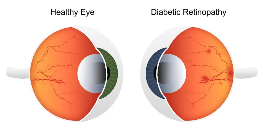
What is Diabetic Retinopathy?
Diabetic retinopathy is an eye condition caused by damage to the tiny blood vessels in the retina due to high blood sugar levels from diabetes. The longer you’ve had diabetes, the higher your risk of developing this condition. Both types of diabetics are at risk. The good news is that most cases can be prevented with early detection and treatment. That’s why it’s especially important to have a comprehensive eye exam at least once a year.
What causes diabetic retinopathy? When blood sugar is poorly controlled, blood vessels in the retina may leak fluid or blood and leave deposits there. This in turn causes swelling. In some people, however, it’s caused by the growth of abnormal new blood vessels on the retina’s surface.
PRK
Early Stages
Background Diabetic Retinopathy (BDR) is an early stage, more like a warning sign. It indicates that serious damage to the eye may be starting. BDR can occur at any time after the onset of diabetes. It often has no visual symptoms, but is usually caught in routine eye exams. Generally, patients at this stage are closely monitored.
The condition may advance to pre-proliferative diabetic retinopathy. At this point, the retina swells. The leaking blood vessels must be sealed by performing a laser photocoagulation on the retina.
Advanced Stages
Proliferative Diabetic Retinopathy (PDR) happens when fragile, abnormal blood vessels grow along the surface of the retina and across the vitreous (the clear jelly inside the eye). Should they break or even leak, severe vision loss or blindness can occur. Alternatively, fluid may leak into the center of the macula. The macula is the part of the eye that allows you to see sharp images straight ahead. Patients with PDR should receive scatter laser photocoagulation (aka pan-retinal laser photocoagulation) to seal the vessels as soon as possible after diagnosis.
Avoiding Blindness
Left too long without treatment, the vessels can bleed into the vitreous. This may also cause the retina to detach, potentially causing blindness. In either case, you will need a vitrectomy. This is an operation to remove the hemorrhage, as well as the broken blood vessels. It’s important to remember that not all diabetics will lose their vision. You can reduce or eliminate risks by controlling your blood sugar and blood pressure. This can be accomplished by eating right, getting regular exercise, and having your eyes examined at least once a year.
Treating Diabetic Retinopathy at LaserVue
If you are diagnosed with diabetic retinopathy, don’t put off treatment. Early detection and early treatment will save your sight. Want more information? Interested in scheduling an appointment with with our San Francisco Bay area eye doctors? Contact us or give us a call at 1-800-527-3745
FAQ’s
What is diabetic retinopathy?
Diabetic retinopathy occurs when chronically high blood sugar levels damage the small blood vessels in the retina, the light-sensitive tissue at the back of the eye. This damage can cause the vessels to swell, leak fluid or blood, or lead to the growth of abnormal new blood vessels, ultimately affecting vision.
What are the symptoms?
In its early stages, diabetic retinopathy often has no noticeable symptoms. As the condition progresses, symptoms may include:
- Blurred or fluctuating vision
- Seeing dark spots, floaters (small spots “floating” in your vision), or empty/dark areas in your field of vision
- Impaired color vision
- Difficulty seeing at night
- Sudden vision loss
Can it be treated?
Yes, it can be treated, but vision loss that has already occurred may be irreversible. Treatment options depend on the stage and severity of the condition and may include:
- Careful management of blood sugar, blood pressure, and cholesterol.
- Laser therapy to seal leaking blood vessels or prevent new abnormal ones from growing.Medication injections into the eye to reduce swelling.
- Surgery (e.g., a vitrectomy) for advanced cases involving significant bleeding or scar tissue.
What types of laser therapy are used for diabetic retinopathy?
The two primary types of laser therapy used to treat diabetic retinopathy are focal laser treatment and panretinal photocoagulation (PRP), also known as scatter laser treatment.
Focal Laser Treatment (Focal Photocoagulation)
Purpose: This treatment is primarily used for diabetic macular edema (DME), a condition where fluid leaks into the macula (the central part of the retina responsible for sharp, detailed vision).
Mechanism: The ophthalmologist identifies specific leaking microaneurysms (small bulges in the blood vessels) or areas of diffuse leakage, often using fluorescein angiography imaging. A focused laser is then used to create small burns that seal these specific leaking vessels, which reduces the fluid buildup and helps stabilize or improve vision.
Application: It is typically used when the macula is swollen but the center is not yet severely damaged.
Panretinal Photocoagulation (PRP) (Scatter Laser Treatment)
Purpose: PRP is the standard treatment for proliferative diabetic retinopathy (PDR), an advanced stage of the disease characterized by the growth of abnormal, fragile new blood vessels on the retina.
Mechanism: The laser is used to apply hundreds of small burns to the peripheral areas of the retina, away from the macula. This process reduces the overall oxygen demand of the retina, which in turn causes the abnormal new blood vessels to shrink and often prevents them from regrowing, thus reducing the risk of severe bleeding (vitreous hemorrhage) and retinal detachment.
Application: The main goal is to preserve existing central vision and prevent severe vision loss, rather than to restore vision that has already been lost.Newer TechniquesMore modern approaches often use advanced laser delivery systems such as pattern scanning lasers (e.g., PASCAL laser) or micropulse laser therapy, which offer advantages such as less pain, less collateral tissue damage, and faster treatment sessions compared to conventional single-spot lasers. These newer techniques use shorter laser pulses or different wavelengths to achieve the desired therapeutic effect with less impact on surrounding healthy tissue.
Who is at risk of developing it?
Anyone with any type of diabetes—type 1, type 2, or gestational—is at risk. The primary risk factors for developing and worsening the condition include:
- The duration of time a person has had diabetes.
- Poor control of blood sugar levels (hyperglycemia).
- High blood pressure and high cholesterol.
- Smoking.
- Pregnancy.
How is it diagnosed?
Diabetic retinopathy is typically diagnosed through a comprehensive dilated eye exam, during which an eye doctor can view the back of the eye to look for signs of damage. Specialized tests like retinal imaging or fluorescein angiography may also be used to evaluate blood flow and swelling in the retina.
Can it be prevented?
While it cannot always be entirely prevented, the risk of developing or worsening diabetic retinopathy can be significantly lowered by:
- Maintaining strict control of blood sugar, blood pressure, and cholesterol levels.
- Adopting a healthy lifestyle, including diet and exercise.
- Having regular, annual dilated eye exams, as recommended by healthcare providers.
- For more information and resources, visit the National Eye Institute or the American Diabetes Association.
Ready to SEE a Change?
Helping you to achieve your personal best vision is our mission at LaserVue Eye Center. Wondering where to start?
Please complete the fields on the right to schedule a free consultation and discuss your options with our eye doctors! You can also Call 1-800-527-3745 to speak with our staff.
