

Cataract surgery is the most commonly performed surgical procedure in the United States, with more than 4 million operations completed each year, according to the American Academy of Ophthalmology’s 2025 clinical summary. The serious complication rate sits below 1.5% at experienced centers. And yet patients in Santa Rosa and the North Bay arrive at consultations asking the same anxious question: is it actually safe? That question deserves a direct, evidence-based answer, not reassurance, but data. This post walks through the clinical evidence on cataract surgery outcomes, the real risks, how surgeons manage them, and what to specifically look for when choosing a provider in the Santa Rosa area.
TL;DR — What the evidence shows:
- Cataract surgery has a 95–98% success rate in improving vision, making it one of the safest elective procedures in medicine, according to AAO 2025 data.
- Serious complications occur in fewer than 1.5% of procedures at experienced centers.
- Most “complications” discussed online are manageable, temporary, or treatable, not permanent vision loss.
- The surgical technology used in Santa Rosa and the North Bay is the same FDA-cleared equipment used at major academic medical centers.
- Delaying cataract surgery carries its own risk, including falls, driving accidents, and accelerated functional decline.
What Cataract Surgery Actually Does
A cataract is the progressive clouding of the eye’s natural crystalline lens. The lens sits behind the iris and focuses light onto the retina. When it clouds, light scatters rather than focuses, producing blurry vision, glare, halos around lights, faded color perception, and difficulty with contrast. No eye drop, supplement, or lifestyle change reverses it. The only treatment is surgical removal of the clouded lens and replacement with a clear artificial intraocular lens (IOL).
The procedure is performed under topical anesthesia (eye drops, not general anesthesia) and takes 10–20 minutes. The surgeon makes a small incision, typically 2–3 mm, uses ultrasound energy to break up the clouded lens (a technique called phacoemulsification), removes the pieces by suction, and inserts a folded artificial IOL through the same incision. The IOL is positioned within the eye, in the lens capsule. No stitches are required in most cases.
What “Safe” Means Clinically
When ophthalmologists call cataract surgery safe, they mean two things. First, the probability of a serious adverse event is low, consistently documented at or below 1.5% across large population studies. Second, the risk-benefit ratio strongly favors surgery for patients with visually significant cataracts: the functional gains (improved visual acuity, contrast sensitivity, color perception, and reduced glare) are substantial and durable, and the risks of delay are real and accumulating.
That clinical definition differs from “zero risk,” which does not exist in any surgical context. The honest answer to “Is it safe?” is it is among the safest surgeries performed on any part of the human body, with outcome data from tens of millions of procedures globally.
The Real Complication Rate: What the Data Shows
Outcome data for cataract surgery are among the most robust in all of surgery because the procedure is so common. Large population studies, national registries, and clinical trial data all converge on consistent numbers.
Posterior Capsule Rupture
The most discussed intraoperative complication is posterior capsule rupture, a tear in the membrane that holds the lens. This occurs in approximately 0.5–2% of procedures, according to data from the European Registry of Quality Outcomes for Cataract and Refractive Surgery (EUREQUO). In most cases, the surgeon manages it during the operation, and a different IOL is used, with a good visual outcome. In a small subset of rupture cases, referral to a retinal specialist is needed. Permanent serious vision loss directly attributable to posterior capsule rupture is rare.
Posterior Capsule Opacification (PCO)
PCO, sometimes called a “secondary cataract,” is not a complication of surgery itself but a natural biological response. Lens epithelial cells that remain after surgery can migrate across the posterior capsule, causing cloudiness weeks to years later. It occurs in roughly 20% of patients within 5 years. Treatment is a 5-minute outpatient laser procedure (YAG capsulotomy) that restores clear vision immediately. It is not re-surgery; it does not require anesthesia or an incision.
Infection (Endophthalmitis)
Endophthalmitis, an infection inside the eye, is the most feared complication of cataract surgery because, if left untreated, it can cause permanent vision loss. The rate at centers using current antiseptic and prophylactic antibiotic protocols is approximately 0.03–0.05%, meaning 3–5 cases per 10,000 procedures. Centers using intracameral antibiotic injection at the close of surgery, the current standard of care endorsed by the ASCRS, achieve rates at the lower end of that range.
Prompt recognition and treatment are critical. Patients who report sudden pain, severe redness, or vision deterioration within two weeks of surgery require same-day evaluation.
Cystoid Macular Edema (CME)
CME is swelling of the central retina (macula) following surgery, caused by an inflammatory response. It is the most common cause of reduced vision after otherwise uncomplicated cataract surgery, occurring in roughly 1–2% of cases. Most resolve with topical anti-inflammatory drops over 4–8 weeks. Persistent CME requires additional treatment but rarely causes permanent, significant vision loss when managed promptly.
Retinal Detachment
Cataract surgery slightly increases the lifetime risk of retinal detachment, particularly in highly myopic (nearsighted) eyes. The increased risk is small, approximately 0.5–1% above baseline over 10 years, and is highest in the first year after surgery. Patients with high myopia are counseled specifically about this risk and instructed to report flashes or floaters immediately.
| Complication | Rate | Typically Manageable? |
|---|---|---|
| Posterior capsule rupture | 0.5–2% | Yes, managed intraoperatively in most cases |
| Posterior capsule opacification (PCO) | ~20% within 5 years | Yes, laser treatment in 5 minutes |
| Endophthalmitis (infection) | 0.03–0.05% | Yes, with prompt diagnosis and treatment |
| Cystoid macular edema (CME) | 1–2% | Yes, resolves with drops in most cases |
| Retinal detachment | 0.5–1% above baseline | Yes, with prompt retinal surgery |
| Serious permanent vision loss | <0.5% | Requires urgent intervention |
Data sources: AAO 2025, ESCRS EUREQUO Registry, ASCRS 2024 Clinical Survey
What Increases Your Risk, and What Doesn’t
Not every patient carries the same risk profile. Understanding which factors matter helps patients and surgeons make informed decisions about timing, technique, and IOL selection.
Factors That Genuinely Affect Risk
Cataract maturity. A dense, mature cataract requires more ultrasound energy to break up, increasing operative time and the risk of corneal swelling and posterior capsule stress. Earlier surgery, before the cataract reaches maximum density, is consistently associated with better outcomes. Waiting until vision is severely impaired is not clinically advantageous.
Pre-existing ocular conditions. Glaucoma, macular degeneration, diabetic retinopathy, pseudoexfoliation syndrome, and a history of uveitis each require modified surgical planning and affect outcome expectations. These are not contraindications; they require disclosure and adjusted technique.
High myopia. Eyes with very long axial length (high nearsightedness) have a higher baseline risk of retinal detachment and some technical challenges with IOL power calculation. Experienced surgeons manage this routinely.
Surgeon experience and volume. Complication rates correlate with surgical volume. A surgeon performing fewer than 50 procedures per year has a significantly higher complication rate than one performing 400+ procedures, according to a population-level analysis of the ESCRS registry. Volume is a reasonable and appropriate question to ask any provider.
Zonular integrity. The zonules are the fibers that hold the lens capsule in place. Pseudoexfoliation syndrome, trauma history, or genetic conditions affecting connective tissue can weaken them, creating technical complexity. This is identified pre-operatively and managed with capsular tension rings or a modified technique.
Factors That Don’t Affect Risk (But Worry Patients)
Age alone. Cataract surgery is routinely performed successfully in patients in their 80s and 90s. Age by itself is not a risk factor for surgical complications, though age often correlates with comorbidities that are.
Nervousness or anxiety. Patient anxiety does not increase complication rates. Most centers offer mild oral sedation for anxious patients. The procedure is performed under topical drops, and patients are awake but comfortable.
Having surgery on both eyes. Sequential bilateral cataract surgery, typically with 1–2 weeks between eyes, is standard practice and does not compound risk. Simultaneous same-day bilateral surgery is performed at some centers but is not a universal standard of care for cataracts, unlike for refractive procedures.
Choosing Between IOL Types: The Decision That Shapes Your Vision
Cataract surgery removes the lens and replaces it with a new one. The IOL choice determines your vision after surgery. This is the most consequential decision in the process, and online information about it is often confusing or incomplete.
Monofocal IOLs
The standard IOL corrects for one focal distance, typically distance vision. Most patients with monofocal IOLs achieve good distance vision without glasses but use reading glasses for near tasks. Monofocal lenses are covered by Medicare and most insurance plans. They have the longest track record and the highest optical quality for their focal point.
Multifocal and Extended Depth of Focus (EDOF) IOLs
Premium IOLs, multifocal and EDOF designs, aim to reduce spectacle dependence by providing multiple focal zones. They are not covered by standard insurance plans; the premium is paid out of pocket. Trade-offs include higher rates of halos and glare at night compared to monofocal IOLs, particularly in the early adaptation period. Neuroadaptation over 3–6 months reduces these symptoms in most patients. Candidates with macular pathology or irregular astigmatism are typically not good candidates for premium IOLs, as these conditions limit the contrast sensitivity required by premium designs.
Toric IOLs
Toric IOLs correct astigmatism simultaneously with the cataract correction. They are available in both monofocal and multifocal designs. For patients with pre-existing corneal astigmatism of 0.75 diopters or more, a toric IOL typically produces better unaided visual acuity than a spherical IOL. Accurate preoperative biometry and proper IOL alignment at surgery are essential for optimal toric outcomes.
| IOL Type | Focal Range | Covers Astigmatism | Insurance Coverage | Best For |
|---|---|---|---|---|
| Monofocal (standard) | Single distance | No | Yes (Medicare/most plans) | Patients comfortable with reading glasses |
| Monofocal toric | Single distance | Yes | Partial | Patients with astigmatism accepting reading glasses |
| EDOF | Distance + intermediate | Optional (toric versions available) | No (premium) | Active patients; computer users; mild night vision sensitivity |
| Multifocal | Distance + nearness | Optional | No (premium) | Patients prioritizing spectacle independence |
| Light-adjustable lens (LAL) | Adjusted post-op | No | No (premium) | Patients prioritizing precision: post-refractive surgery eyes |
The Risk of Not Having Surgery
Cataract surgery discussions focus on operative risk. The risk of delay is discussed far less often, and it is real.
A prospective study published in JAMA Ophthalmology found that older adults with visually significant cataracts have a 130% higher fall rate than age-matched peers with normal vision. Falls in adults over 65 are the leading cause of injury-related death in the United States, according to the CDC. Reduced contrast sensitivity and glare from cataracts also impair driving performance; a 2024 analysis in Ophthalmology found cataract-associated driving impairment comparable to a blood alcohol level of 0.08%.
Beyond safety, there is a functional quality of life. Reading, cooking, recognizing faces, watching television, and navigating unfamiliar environments all become progressively more difficult as cataracts advance. Surgery restores function quickly and durably.
“The data on delay is unambiguous: for patients with visually significant cataracts, the risks of waiting, falls, accidents, functional decline, social withdrawal, consistently exceed the risks of a well-performed surgery.” — American Academy of Ophthalmology, Preferred Practice Pattern: Cataract in the Adult Eye, 2025
What Makes Cataract Surgery Safe: The Technology Side
The safety profile of modern cataract surgery is not accidental. It reflects decades of technology development in three areas.
Phacoemulsification
Phacoemulsification, ultrasound-based lens fragmentation, replaced the older extracapsular technique in the 1980s and 1990s. The shift reduced incision size from 10–12 mm to 2–3 mm, eliminated the need for sutures, reduced operative time, and dramatically reduced the rate of surgically induced astigmatism and wound-related complications. All cataract surgery at established North Bay centers uses phacoemulsification.
Femtosecond Laser-Assisted Cataract Surgery (FLACS)
FLACS uses a femtosecond laser to perform the incisions, lens fragmentation, and capsulorhexis (the circular opening in the lens capsule) with computer-guided precision. The technology reduces variability in the most technique-sensitive steps of the procedure. Evidence on whether FLACS produces meaningfully better visual outcomes than skilled manual phacoemulsification in routine cases is mixed, but it offers advantages in specific situations, dense cataracts, toric IOL alignment, and post-refractive surgery eyes. Not all Santa Rosa-area centers offer FLACS; it is reasonable to ask whether it is available and, if so, under what circumstances it would be recommended.
Advanced Biometry
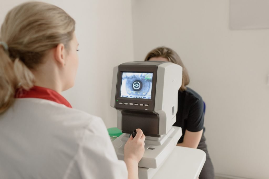
IOL power calculation has been revolutionized by optical coherence-based biometry (devices such as the Zeiss IOLMaster 700 and Haag-Streit Lenstar). These instruments measure axial length, corneal curvature, anterior chamber depth, and lens thickness with sub-micron precision, feeding modern IOL formulas (Barrett Universal II, Kane, and Hill-RBF) that achieve target refraction within 0.50 diopters in over 90% of cases. Poor biometry is the leading cause of refractive surprise after cataract surgery, an avoidable outcome at centers using current equipment.
How to Evaluate a Cataract Surgeon in Santa Rosa or the North Bay
The difference between a good outcome and a difficult one is often less about luck and more about who you choose and how they prepare.
| What to Evaluate | What to Look For | Red Flag |
|---|---|---|
| Surgeon credentials | Board-certified ophthalmologist; fellowship in anterior segment or comprehensive ophthalmology | Optometrist-performed surgery; unlisted credentials |
| Annual surgical volume | 300+ cases/year at the practice | Vague answer; fewer than 50 personally |
| Biometry equipment | IOLMaster 700, Lenstar, or equivalent optical biometry | “State-of-the-art” without naming the device |
| IOL options offered | Full range including toric and premium IOLs | Only standard monofocal offered |
| FLACS availability | Available for appropriate cases; explained clearly | Pushed aggressively on all patients regardless of complexity |
| Infection protocol | Intracameral antibiotic at close of surgery | Topical drops only as prophylaxis |
| Co-management clarity | Clear on who handles post-op if complications arise | Unclear referral pathway |
| Premium IOL counseling | Trade-offs of premium vs. standard explained honestly | Premium IOLs sold without discussing halos/glare |
Cataract Surgery and Specific Health Conditions
Diabetes
Diabetic patients have higher baseline rates of macular edema and retinal complications. Pre-operative retinal evaluation is recommended. Outcomes are generally excellent when retinopathy is not advanced, but realistic expectations should be discussed.
Glaucoma
Cataract surgery can lower intraocular pressure in some glaucoma patients, a secondary benefit. For patients with advanced glaucoma, combined cataract and glaucoma surgery (with MIGS, minimally invasive glaucoma surgery devices) may be considered. IOL selection requires careful discussion, as visual field loss can affect candidacy for premium IOLs.
Macular Degeneration
Cataract surgery does not cause or accelerate AMD. It can be performed safely in AMD patients, though premium multifocal IOLs are contraindicated because compromised macular function limits their effectiveness. Standard monofocal IOLs improve the optical clarity of the eye even when retinal disease persists.
Prior LASIK or Refractive Surgery
Patients who have had LASIK, PRK, or RK (radial keratotomy) require a modified IOL power calculation. Standard biometry formulas underperform in post-refractive eyes; practices using corneal topography-adjusted formulas and intraoperative wavefront aberrometry (ORA) achieve better refractive outcomes in this population. If you have had prior corneal refractive surgery, confirm that your surgeon has a specific protocol for post-refractive IOL calculation.
Book Your Comprehensive Cataract Evaluation in Santa Rosa Today
We follow the same clinical standards used at leading academic centers, right here in your community. You’ll leave knowing exactly what’s best for your vision, not just what you read online.

Santa Rosa & North Bay | Board-certified ophthalmologists | Advanced laser & biometry technology
Frequently Asked Questions
How safe is cataract surgery for someone over 75?
Cataract surgery is routinely performed safely in patients in their 70s, 80s, and 90s. Age alone is not a risk factor. The relevant factors are ocular health (including AMD, glaucoma, or other conditions), systemic factors that affect healing, and the surgeon’s experience with the specific complexity of the case. Outcomes data from population-level studies include large numbers of older patients with excellent results.
Will I need glasses after cataract surgery?
It depends on the IOL chosen. Standard monofocal IOLs correct for distance; most patients use reading glasses for near tasks. Premium multifocal or EDOF IOLs reduce dependence on glasses for most distances but do not guarantee spectacle independence and carry higher rates of halos and glare. Toric IOLs correct pre-existing astigmatism. The right choice depends on your lifestyle priorities, visual demands, and ocular health.
Does Medicare cover cataract surgery in Santa Rosa?
Medicare Part B covers cataract surgery when it is medically necessary, defined as a cataract that causes visual impairment and affects daily function. The standard monofocal IOL is covered. The premium portion of upgraded IOLs (toric, multifocal, EDOF, and light-adjustable lenses) is an out-of-pocket cost for patients. Most supplemental Medigap policies cover the copays and deductibles for the covered portion.
How long does cataract surgery last?
The IOL implanted during cataract surgery is permanent. It does not expire, degrade, or require replacement. The most common post-surgical development is posterior capsule opacification (PCO), a natural clouding of the membrane behind the IOL, which occurs in roughly 20% of patients within 5 years and is corrected with a quick, painless in-office laser treatment.
Can I have cataract surgery if I’ve previously had LASIK?
Yes, but with an important caveat: prior LASIK changes the corneal shape in ways that can cause standard IOL power calculations to underestimate or overestimate the needed lens power. Practices that use corneal topography-adjusted formulas and intraoperative aberrometry (ORA system) achieve better refractive outcomes in post-LASIK eyes. Confirm your provider has a specific protocol for this before proceeding.
What is the difference between traditional and laser cataract surgery?
Traditional cataract surgery uses a manual blade for incisions and capsulorhexis, then phacoemulsification to remove the lens. Laser-assisted cataract surgery (FLACS) uses a femtosecond laser to create incisions and fragment the lens with computer-guided precision. Both achieve excellent outcomes in routine cases. FLACS offers advantages in specific complex situations. The decision should be driven by clinical indication, not by upselling.
See also: Toric Lenses in Santa Rosa: Correcting Astigmatism During Cataract Surgery | Cataract Surgery Recovery in Santa Rosa: What to Expect Week by Week
About LaserVue Eye Center
LaserVue Eye Center has been the Bay Area’s and Sonoma County’s trusted destination for LASIK, cataract surgery, and premium vision correction since 1996. Our board-certified ophthalmologists have performed thousands of procedures across our San Francisco and Santa Rosa locations, combining advanced surgical technology with the individualized care that brings patients back and earns referrals from their neighbors, colleagues, and physicians. Learn more about our cataract surgery program and meet our surgical team.